黑色素瘤相关抗原抗体
产品名称: 黑色素瘤相关抗原抗体
英文名称: CD63
产品编号: 1523
产品价格: null
产品产地: 上海
品牌商标: 雅吉
更新时间: null
使用范围: WB ELISA IHC-P IHC-F Flow-Cyt ICC IF
- 联系人 :
- 地址 : 上海市闵行区元江路5500号第1幢5658室
- 邮编 :
- 所在区域 : 上海
- 电话 : 158****3937
- 传真 : 021-34661275
- 邮箱 : yajikit@163.com
| 中文名称 | 黑色素瘤相关抗原抗体 |
| 别 名 | Lysosomal associated membrane protein 3; CD63 antigen; CD63 antigen melanoma 1 antigen; CD63 molecule; granulophysin; LAMP 3; LAMP3; lysosome associated membrane glycoprotein 3; CD 63; CD63; CD63 antigen (melanoma 1 antigen); CD63_HUMAN; gp55; granulophysin; LAMP-3; LIMP; Lysosomal-associated membrane protein 3; Melanoma-associated antigen ME491; NGA; Ocular melanoma-associated antigen; PTLGP40; Tetraspanin-30; Tspan-30; Mast cell antigen AD1; ME491; melanoma 1 antigen; Melanoma associated antigen ME491; Melanoma associated antigen MLA1; MGC72893; MLA 1; MLA1; ocular melanoma associated antigen; OMA81H; Tetraspanin 30; Tspan 30; TSPAN30; Tumor biomarkers; Platelets markers. |
研究领域 肿瘤 心血管 免疫学 细胞类型标志物
抗体来源 Rabbit
克隆类型 Polyclonal
交叉反应 Human,
产品应用 WB=1:500-2000 ELISA=1:500-1000 IHC-P=1:100-500 IHC-F=1:100-500 Flow-Cyt=1μg/Test ICC=1:100-500 IF=1:100-500 (石蜡切片需做抗原修复)
not yet tested in other applications.
optimal dilutions/concentrations should be determined by the end user.
分 子 量 26kDa
细胞定位 细胞浆 细胞膜
性 状 Liquid
浓 度 1mg/ml
免 疫 原 KLH conjugated synthetic peptide derived from human CD63:101-200/238
亚 型 IgG
纯化方法 affinity purified by Protein A
储 存 液 0.01M TBS(pH7.4) with 1% BSA, 0.03% Proclin300 and 50% Glycerol.
保存条件 Shipped at 4℃. Store at -20 °C for one year. Avoid repeated freeze/thaw cycles.
PubMed PubMed
产品介绍 The protein encoded by this gene is a member of the transmembrane 4 superfamily, also known as the tetraspanin family. Most of these members are cell-surface proteins that are characterized by the presence of four hydrophobic domains. The proteins mediate signal transduction events that play a role in the regulation of cell development, activation, growth and motility. This encoded protein is a cell surface glycoprotein that is known to complex with integrins. It may function as a blood platelet activation marker. Deficiency of this protein is associated with Hermansky-Pudlak syndrome. Also this gene has been associated with tumor progression. The use of alternate polyadenylation sites has been found for this gene. Alternative splicing results in multiple transcript variants encoding different proteins.
Function:
This antigen is associated with early stages of melanoma tumor progression. May play a role in growth regulation.
Subcellular Location:
Cell membrane; Multi-pass membrane protein. Lysosome membrane; Multi-pass membrane protein. Late endosome membrane; Multi-pass membrane protein. Note=Also found in Weibel-Palade bodies of endothelial cells. Located in platelet dense granules.
Tissue Specificity:
Dysplastic nevi, radial growth phase primary melanomas, hematopoietic cells, tissue macrophages.
Similarity:
Belongs to the tetraspan
| 产品图片 |
 Sample:
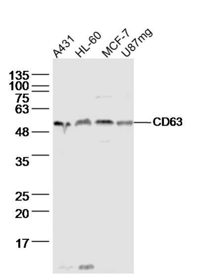
A431 cell(human) Lysate at 40 ug HL-60 cell(human) Lysate at 40 ug MCF-7 cell(human) Lysate at 40 ug U87mg cell(human)Lysate at 40 ug Primary: Anti- CD63 (bs-1523R)at 1/300 dilution Secondary: IRDye800CW Goat Anti-Rabbit IgG at 1/20000 dilution Predicted band size: 26kD Observed band size: 53 kD 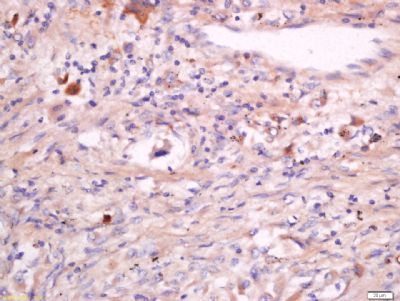 Tissue/cell: human lung carcinoma; 4% Paraformaldehyde-fixed and paraffin-embedded;
Antigen retrieval: citrate buffer ( 0.01M, pH 6.0 ), Boiling bathing for 15min; Block endogenous peroxidase by 3% Hydrogen peroxide for 30min; Blocking buffer (normal goat serum,C-0005) at 37℃ for 20 min; Incubation: Anti-CD63 Polyclonal Antibody, Unconjugated(bs-1523R) 1:200, overnight at 4°C, followed by conjugation to the secondary antibody(SP-0023) and DAB(C-0010) staining 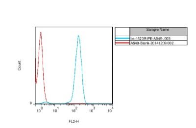 cell: A549 cells
Incubation: Avoid light, at 4°C for 60 minutes. Blank control A549 cells(red line),5X10^5/ml, at 4°C for 60 minutes. primary antibody: abbit Anti-CD63 antibody (bs-1523R-PE,Blue line) ,1:50, at 4°C for 60 minutes. 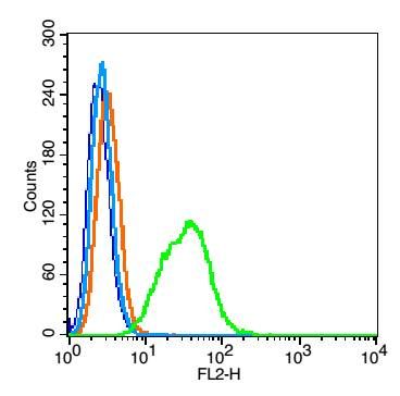 Blank control: Hep G2 cells(blue).
Primary Antibody:Rabbit Anti- CD23 antibody(bs-1523R), Dilution: 1μg in 100 μL 1X PBS containing 0.5% BSA; Isotype Control Antibody: Rabbit IgG(orange) ,used under the same conditions ); Secondary Antibody: Goat anti-rabbit IgG-PE(white blue), Dilution: 1:200 in 1 X PBS containing 0.5% BSA. Protocol The cells were fixed with 2% paraformaldehyde (10 min) , then permeabilized with 90% ice-cold methanol for 30 min on ice. Primary antibody (bs-1523R,1μg /1x10^6 cells) were incubated for 30 min on the ice, followed by 1 X PBS containing 0.5% BSA + 1 0% goat serum (15 min) to block non-specific protein-protein interactions. Then the Goat Anti-rabbit IgG/PE antibody was added into the blocking buffer mentioned above to react with the primary antibody at 1/200 dilution for 30 min on ice. Acquisition of 20,000 events was performed. |